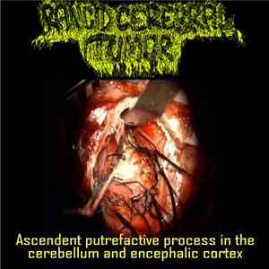
Rancid Cerebral Tumor – Ascendent Putrefactive Process In The Cereberebellum And Encephalic Cortex. Label: Rectal Purulence – RP 008.
Rancid Cerebral Tumor. Ascendent Putrefactive Process In The Cereberebellum And Encephalic Cortex (CD, Mini, B/card). Rancid Cerebral Tumor, Pineal Tumor Surgery - 2 Ways To Develop Mesocortical Intra-Axial Neoplasias In The 3th Ventricule With Clinical Manifestation Of Intracranial Hypertension Causing Massive Thromboembolia (CDr, Ltd, Num).
Rancid cerebral tumor (full album) 2015. July 30, 2017 ·. Rancid Cerebral Tumor - Steatosis Of The Epithelia With Parcial Adipocerous Dermatopyosis youtube. 30 July 2017 ·.
A cerebral infarction is an area of necrotic tissue in the brain resulting from a blockage or narrowing in the arteries supplying blood and oxygen to the brain. The restricted oxygen due to the restricted blood supply causes an ischemic stroke that can result in an infarction if the blood flow is not restored within a relatively short period of time. The blockage can be due to a thrombus, an embolus or an atheromatous stenosis of one or more arteries.
The symptoms of a tumor of the cerebellum are of gradual onset, and unfortunately their benign or malignant nature cannot be predicted. There are two types of symptoms associated with a tumor of the cerebellum. The first type includes symptoms related to the cerebellum itself, which are grouped under the term cerebellar syndrome. They cover: headache during increased physical activity, which are typically stronger in the morning; nausea or vomiting (projectile); and blurred vision or double vision. These symptoms are inconsistent and may not all be present at once, depending on the location of the tumor. The diagnosis of a cerebellar tumor is based on the clinical signs. A CT scan or MRI will highlight the injury, and depending on the features found in imaging, the nature of it.
The cerebral cortex is connected to various subcortical structures such as the thalamus and the basal ganglia. Most sensory information is routed to the cerebral cortex via the thalamus. Olfactory information, however, passes through the olfactory bulb to the olfactory cortex, bypassing the thalamus. Buried deep in the white matter of the cerebral cortex are interconnected subcortical masses of cerebral gray matter called basal nuclei (or basal ganglia) that are involved in motor control. The basal nuclei receive input from the substantia nigra of the midbrain and motor areas of the cerebral cortex and send signals back to both of these locations. Motor Cortex Map. The majority of neurons in the motor cortex project to the spinal cord synapse on interneuron circuitry in the spinal cord
In the cerebral cortex isolated sensory, motor, and association areas (Fig. 1. 1). Brodman's cytoarchitectonic fields. The cortical ends of the analyzers have their topography - local location in certain areas of the cerebral cortex. They are called sensory areas of the cerebral cortex. The cortical ends of the analyzers of different sensory systems overlap. In the cortex of the parietal lobe (fields 5 and 7, see Figure 1. 1), where the conduction pathways of sensitivity also terminate, a more complex analysis is carried out: localization of irritation, discrimination, stereo- religion. In case of damage to the cortex, the functions of the distal parts of the extremities, especially the hands, are especially severely impaired.
The cerebral cortex (plural cortices), also known as the cerebral mantle, is the outer layer of neural tissue of the cerebrum of the brain, in humans and other mammals. It is separated into two cortices, by the longitudinal fissure that divides the cerebrum into the left and right cerebral hemispheres. The two hemispheres are joined beneath the cortex by the corpus callosum. The cerebral cortex is the largest site of neural integration in the central nervous system.
 TUMOR - splattered human goulash 10" download flac
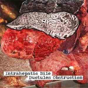 Biliary Cirrhosis - Intrahepatic Bile Ductules Obstruction download flac
 Vulgar Disease - Rectal Anarchy download flac
 Bud Melvin - Business Card CD Single download flac
 Insidious Squelching Penetration - Delectable Rectal Meat download flac
 Anal Cake, Rectal Twat - Untitled download flac